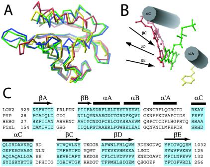
Figure 3.
Structural alignment of four PAS domains. (A) Least-squares superposition: FixL, red; PYP, yellow; HERG, blue; phy3 LOV2, green. (B) Comparison of positions of phy3 LOV2 (green), FixL (red), and PYP (yellow) chromophores within the chromophore-binding pocket of PAS domain. The secondary structural elements are represented schematically as cylinders and arrows. (C) Structure-based alignment of the sequences of these four PAS domains. Residues boxed in blue form secondary structural elements conserved among all four structures. These residues were used to optimize the least-squares fit shown in Fig. 3A. Secondary structure is noted above the alignment: β-strand, arrows; helix, bars.

