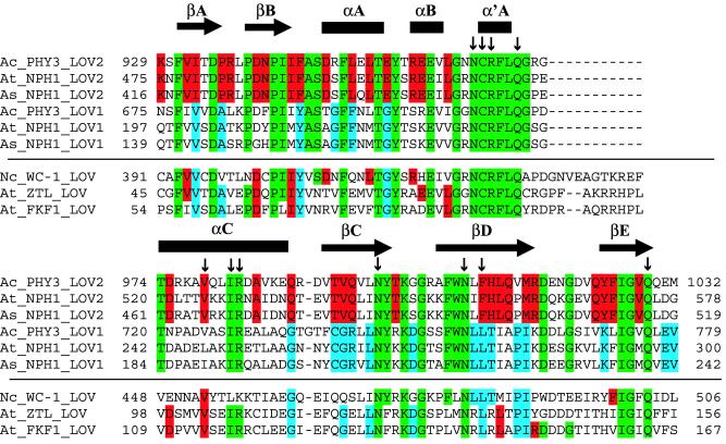
Figure 4.
Alignment of nine LOV-domain sequences and identification of flavin-interacting residues including LOV1 and LOV2 domains from Adiantum phy3 (GenBank accession no. BAA36192), Arabidopsis phototropin (GenBank accession no. AAC01753), oat phototropin (GenBank accession no. AAC05083), Arabidopsis ztl (GenBank accession no. AAF70288), Arabidopsis fkf1 (GenBank accession no. AAF32298), and Neurospora Wc-1 (GenBank accession no. Q01371). LOV2 residues that interact with FMN are marked with vertical arrows. Secondary structure of LOV2 is marked above alignment: β-strand, arrows; helix, bars. Green residues are conserved in all LOV1 and LOV2 domains included in the alignment as well as eight other LOV domains (not included because of space limitations) from rice phototropin (BAA84779), corn phototropin (T01353), rice phototropin-like protein nph1b (GenBank accession no. BAA84779) and Arabidopsis phototropin-like protein npl1 (GenBank accession no. AAC27293). Blue residues are conserved in all LOV1 domains, and red residues are conserved in all LOV2 domains from the above-listed proteins. This color scheme is applied to residues in Wc-1, ztl, and fkf1.

