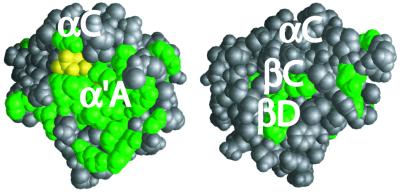
Figure 6.
Space-filling model of phy3 LOV2 showing the surface position of residues conserved in all LOV1 and LOV2 domains (green). Terminal phosphate of FMN is colored yellow. (Left) Model of the conserved face containing the 310-helix α′A. (Right) Model is rotated 180° about a vertical axis in the plane of Fig. 1.

