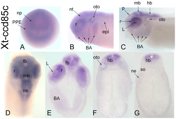
Figure 4.
Expression of a CG17265-related gene, Xt-ccd85c. (A) Diffuse expression of Xt-ccd85c throughout the anterior neural plate and PPE [Anterior view]. (B) At neural tube closure, there is diffuse expression throughout the neural tube, retina dorsal epidermis, otocyst and branchial arches [Side view]. (C) At late tail bud stages the pineal (p), retina, midbrain and hindbrain are stained. There also is weak staining in the lens, otocyst and branchial arches [Side view]. (D) Dorsal view at larval stage showing expression extending into the forebrain. Transverse sections at forebrain (E), hindbrain (F) and caudal hindbrain (G) demonstrate restricted neural expression.

