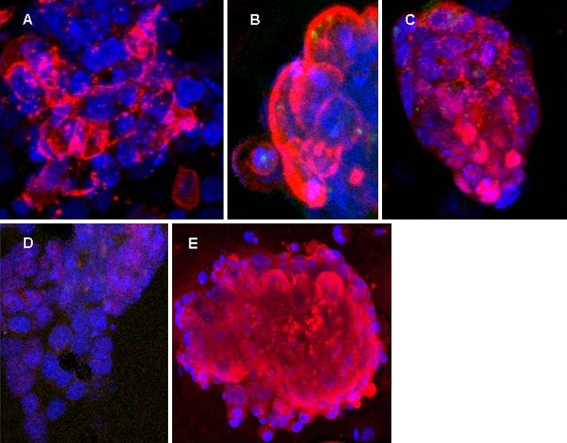
Fig. 2.
Expression and localization of stem cell specific markers in vitrified-warmed ICMs after 48 hours in culture. ICMs were tested for expression of SSEA-1, Sox-2 and Oct-4 using immunoflouresecent staining. Cells were imaged using confocal laser microscopy. (A) ICM stained with SSEA 1 (red signal) and DAPI (blue signal). Staining is detected primarily on ICM cell surface. (B) Enlarged image of ICM cells stained with SSEA-1. (C) Sox-2 staining (red), detected in both nucleus and cytoplasm. (D) Oct-4 staining (red), mostly in the nucleus. MEF and any residual trophectodermal cells were negative for stem cell markers. (E) Oct-4 expression on Day 16 of culture

