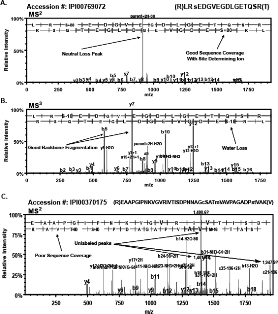
FIGURE 3.
Confident identification of phosphopeptides. The importance of manual validation is illustrated by comparing the spectra of phosphopeptides that were confirmed or rejected. (A) The MS2 scan for a singly phosphorylated peptide (IPI00769072) contains a classic neutral-loss peak with lower intensity sequence coverage of y (lower)- and b (upper)-ions containing the phosphorylation site, determining ion on serine 3. (B) The MS3 scan of the same peptide (IPI00769072) showing improved sequence coverage of y- and b-ions as well as a loss of water from serine 3. (C) The MS2 scan of potential phosphopeptide (IPI00370175) lacks an observable neutral-loss peak, has poor sequence coverage, and has several unidentified, prominent peaks. This spectra illustrates a potential phosphopeptide that passed the probability-filtering and the traditional-score thresholds for the more-stringent filtering criteria but did not satisfy the criteria for manual validation.

