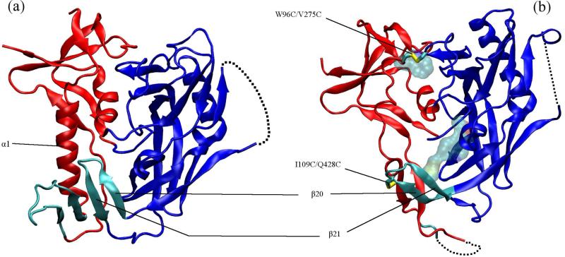
FIG. 1.
(a) CD4-bound (PDB 1GC1) and (b) b12-bound (PDB 2NY7) conformations of gp120. The inner domain is red, outer domain blue and the bridging sheet cyan. In (b) licorice rendering shows the two disulfide bridges (hydrogen atoms not shown) between the inner and outer domain and the residues shown in transparent van der Waals are the other stabilizing mutations (M95W, T257S, S375W, A433M). The dashed lines show the unresolved V4 domain in both structures and also parts of the β2/β3 domain and base of the V1/V2 loop in the b12-bound conformation.

