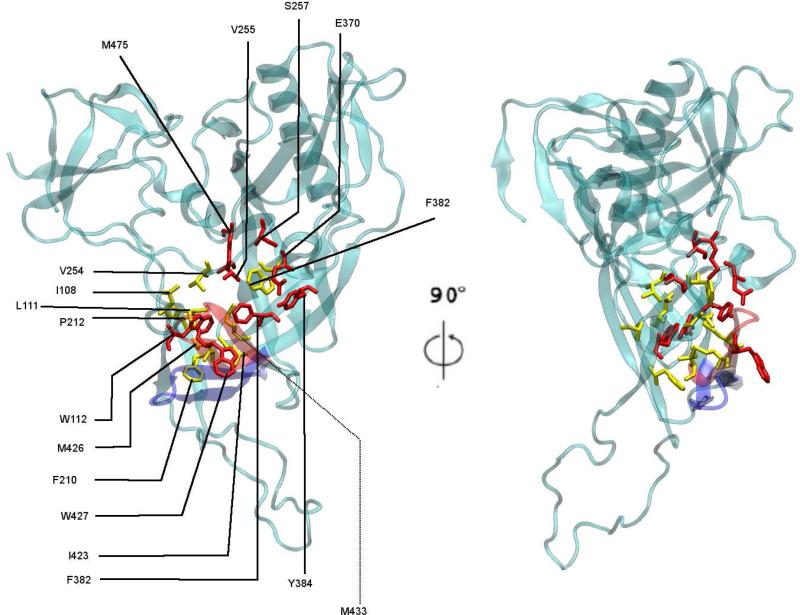
FIG. 4.
Hydrophobic residues in and around the putative F43 pocket in the DS1* structure. Residues in red make up the F43 pocket (5). Residues shown in yellow are hydrophobic residues within 10 Å of residue 382 and taken to be close to the pocket. The position of the β20/β21 domain at the beginning (red) and end (blue) of the DS1* equilibration is shown in cartoon rendering.

