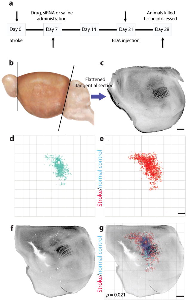
Figure 3.
Quantitative connectional mapping. (a) Timeline of experimental design for all in vivo axonal tracer experiments. Mice received a sham surgery or stroke, followed 1 week later by siRNA or drug delivery. Three weeks later biotinylated dextran amine (BDA) was microinjected into forelimb motor cortex. (b,c) The cortex was then removed, flattened, tangentially cut (b) and stained for cytochrome oxidase (c) and BDA in the same sections. (d,e)The location of BDA-labeled axons was digitally mapped in x, y coordinates relative to the injection site. (d) The quantitative connectional map of forelimb sensorimotor connections in sham-operated animals (n = 5). (e) The quantitative connectional map of forelimb sensorimotor cortex connections after stroke (n = 7). (f) Cytochrome oxidase staining in layer IV identifies the mouse somatosensory body map2 after stroke. (g) Each connectional map was registered to the somatosensory body map from the same brain to produce a group connectional and functional map of cortex. Axonal sprouting was identified when a pattern of cortical connections was precisely mapped and statistically different across treatment conditions. Scale bars, 1 mm.

