
Figure 4.
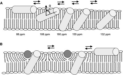
Models of PGLa and magainin 2 helix alignments in oriented lipid bilayers. The PGLa helices are sketched as light gray cylinders, and the magainin 2 helices are viewed along their long axis and shown as dark gray circles. (A) Sketch of how PGLa alone adjusts its alignment to the membrane hydrophobic thickness. Whereas the peptide orients along the membrane surface in thick PC membranes, it spans bilayers made of shorter fatty acyl chains. The approximate 15N chemical shifts of [15N-Ala14]-PGLa are also indicated, as is the definition of the tilt angle, θ, between the membrane normal and the helix long axis. (B) Illustration of how membrane thinning caused by magainin 2 can trigger topological alterations of PGLa in DMPC (but not POPC) membranes (cf. text for details). Small populations or transient additional intermediates (as shown in A) may exist also for PGLa in the presence of magainin 2 but they have been omitted for clarity.