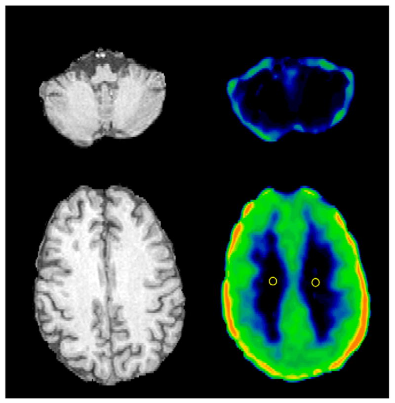
FIGURE 1.

Transverse MR and 18F-FCWAY distribution volume images passing through centrum semiovalis and cerebellum in TLE patient. Negligible binding of tracer in cerebellar white matter and centrum semiovalis and large spillover of 18F-fluoride activity onto cerebellum were observed. Two 6-mm-diameter, circular ROIs used for calculation of VWM with manual approach are displayed. 18F-FCWAY distribution volume image (V/fP) is scaled to maximum of 80 mL/mL.
