
Figure 1.
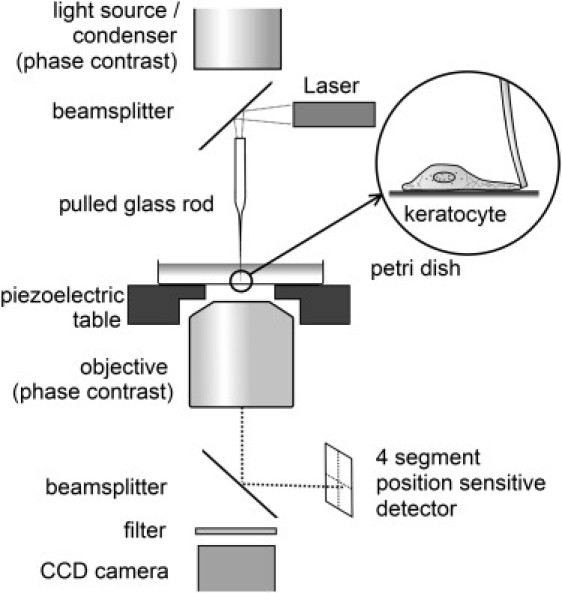
Force microscopy setup for measurement of keratocyte lamellipodial protrusion forces. A laser beam was coupled into a pulled glass rod with a diameter of 5–10 μm at its apex. The thin fiber end of the glass rod was used as a flexible obstacle toward which keratocytes are migrating. The deflection of the fiber was detected on a segmented photo detector.