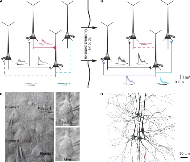
Figure 1.
Evoked emergence of connections following 12 h network stimulation. (A) Recording of layer 5 pyramidal cells (PC) using multiple whole-cell patch-clamp. Direct synaptic connections were examined by eliciting short trains of precisely timed action potentials at 30 Hz. (B) After 12 h of network stimulation by sodium glutamate. The red trace indicates a disappearance, blue and purple traces indicate emergences, grey and black traces indicate stable connections (Le Be and Markram, 2006). (C) Cluster of four cells viewed by infra-red differential interference contrast microscope. (Upper right) Neurons during first patch. (Lower right) Neurons after 12 h of glutamate stimulation. (D) Mesh rendering on camera lucida of anatomical reconstructions of two neurons where a connection appeared after evoking with sodium glutamate. (Red Dots): Putative synaptic contact sites.

