Figure 1.
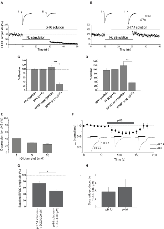
Extracellular acidosis reduces the EPSC. (A) EPSC amplitude was reduced by approximately 70% after stimulation was resumed in the presence of pH6 aCSF. Representative EPSCs show the final baseline response (i) and the first response in pH6 solution (ii). (B) Application of pH7.4 augmented EPSC amplitude following the absence of stimulation, apparent in traces of the final baseline response (i) and the first response evoked in pH7.4 (ii). (C) Initial PFV amplitudes in pH6 and pH7.4 solutions are not significantly different, in contrast to reduction of the initial EPSP slope by pH6. (D) Initial PPR ratios in pH6 and pH7.4 are not significantly different, in contrast to reduction of the initial EPSC amplitude in pH6. (E) Reduction of postsynaptic sensitivity by pH6 is dependent on the concentration of applied glutamate. (F) A reversible modest reduction in amplitude of currents evoked by rapid application of glutamate (3 mM) is produced by pH6 extracellular solution. (G) EPSC amplitude evoked in pH6 is attenuated by γ-DGG (300 μM) to a greater extent than in pH7.4 (p < 0.012). (H) Antagonism of two point concentration-response curves to glutamate (0.3–3 mM) by γ-DGG (300 μM) was not affected by extracellular pH (n = 3, p > 0.65).

