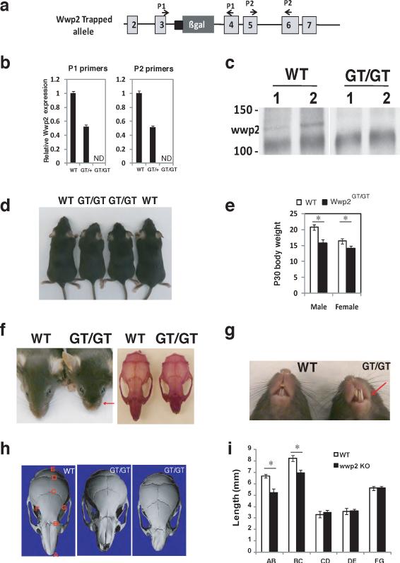
Figure 1. Craniofacial patterning defects are present in Wwp2GT/GT mice.
a) Schematic depicting the position of B-gal insertion into the Wwp2 locus. b) Analysis of Wwp2 transcript levels by qPCR in WT mice (WT), Wwp2GT/+ (GT/+) and Wwp2GT/GT (GT/GT) mice. Values represent means ± s.d. (n=6 for each genotype) c) Immunoprecipitation-Western blot analysis depicting the absence of Wwp2 protein in the Wwp2GT/GT mice. d) Photograph of four-week old male WT and Wwp2GT/GT mice. e) Body weight of male and female WT and Wwp2GT/GT at four-weeks of age. Values represent means ± s.d. (n=6 for each genotype) f) Presence of shortened snout in Wwp2GT/GT mice as shown grossly (left) as well as by alizarin red staining of WT and Wwp2GT/GT skulls (right). g) Photograph depicting misaligned jaw and overgrown of mandibular incisor of Wwp2GT/GT (GT/GT) mice. h) μCT analysis of skulls from WT mice as well as Wwp2GT/GT mice depicting the shortened (middle) or twisted nasal bone (right) in the Wwp2GT/GT mice. i) Quantitative analysis of the distances between the various landmarks in the skulls of four-week old Wt and Wwp2GT/GT mice. Values represent means ± s.d. (n=8 for each genotype) (*p<0.001). Uncropped images of blots are shown in Supplementary Fig.S7.

