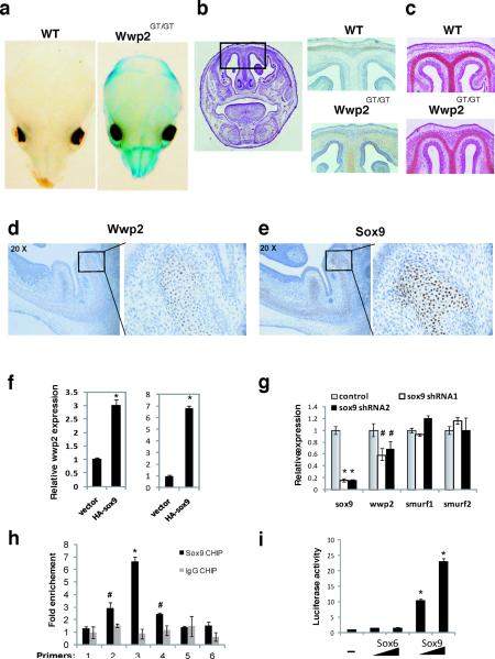
Figure 2. Regulation of Wwp2 expression in the skull.
a) Whole mount staining for β-gal in skulls of WT and Wwp2GT/GT mice. b) Detection of β-gal in histological sections of the skull of Wwp2GT/GT mice but not WT mice via immunostaining. Scale bar, 0.5mm c) Safranin O staining identifies cartilaginous regions of the cranial sections. Scale bar, 0.5mm. Analysis of Wwp2 (d) and Sox9 (e) in the skull of WT mice by in situ hybridization and immunostaining, respectively. Scale bar, 0.5mm. Higher magnification of boxed area in both d) and e) is displayed to right. f) Wwp2 transcript levels were measured by qPCR in ATDC5 cells (left) and C3H10T1/2 cells (right) following infection of cells with control or HA-tagged Sox9-expressing lentivirus. Values represent means ± s.d. (n=3) g) Levels of Sox9, Wwp2, Smurf1 and Smurf2 were evaluated by qPCR following infection of ATDC5 cells with a control lenvtivirus or lentivirus containing Sox9-specific shRNAs. Values represent means ± s.d. (n=3) h) Six separate primer sets (1–6) were used in qPCR reactions to scan the Wwp2 intron region that was bound by Sox9 in chromatin immunoprecipitation experiments in C3H10T1/2 cells that utilized anti-Sox9 (black bars) or IgG control (grey bars) antibody. Values represent means ± s.d. (n=3) i) Increasing amounts of Sox9 but not Sox6 lead to induction of luciferase levels in C3H10T1/2 cells transfected with a luciferase reporter construct that contains the region surrounding the Sox9 binding site in the Wwp2 intron. Values represent means ± s.d. (n=3) (*p<0.01, #p<0.05 compared to empty vector control).

