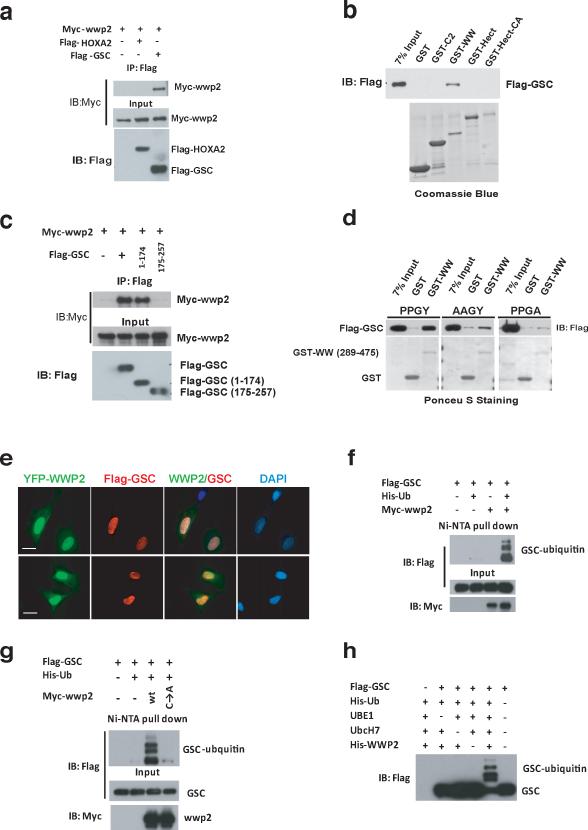
Figure 3. Gsc interacts with and is ubiquitinated by Wwp2.
a) Co-immunoprecipitation experiments were conducted in 293T cells transfected with myc-Wwp2 and flag-Gsc or flag-HoxA2 expression constructs. Wwp2 was immunoprecipitated from cell lysates with anti-flag antibody, followed by Western blot analysis with anti-myc antibody. b) Purified recombinant fragments of Wwp2 were used for in vitro interaction analysis. Western blot analysis with an anti-flag antibody was used to detect Gsc interaction with the various fragments of Wwp2. c) Co-immunoprecipitation experiments were conducted in 293T cells with flag-epitope tagged Gsc deletion mutants and myc-Wwp2 as described in (a). d) Interaction analysis between recombinant WW domain fragments of Wwp2 and wild-type Gsc (PPGY) or Gsc bearing a AAGY or PPGA mutation were analyzed via GST-pull down followed by Western blot analysis. e) Immunofluoresence analysis of ATDC5 chondrocyte cell line transfected with YFP-tagged Wwp2 expression construct and a flag-epitope tagged Gsc expression construct reveals colocalization of these proteins in the nucleus. Scale bar, 10μm f) Analysis of Gsc ubiquitination was performed in 293T cells transfected with flag-Gsc, myc-Wwp2 and His-epitope-tagged ubiquitin. Ubiquitinated flag-Gsc proteins were detected in cells by precipitating ubiquitinated proteins from denatured lysates with Ni-NTA-agarose, followed by Western blot analysis with anti-flag antibody. g) Additional Gsc ubiquitination experiments were conducted as described in f) that utilized either WT Wwp2 or functionally inert Wwp2 harboring a cysteine to alanine mutation. h) In vitro ubiquitination of Gsc was performed using flag-Gsc purified from 293T cells transfected with flag-Gsc. Flag-Gsc was incubated with recombinant Wwp2, His-ubiquitin, UBE1, Ubch7. Reactions were then subjected to Western blot analysis with anti-Flag antibody to detect Gsc proteins. Uncropped images of blots are shown in Supplementary Fig.S7.

