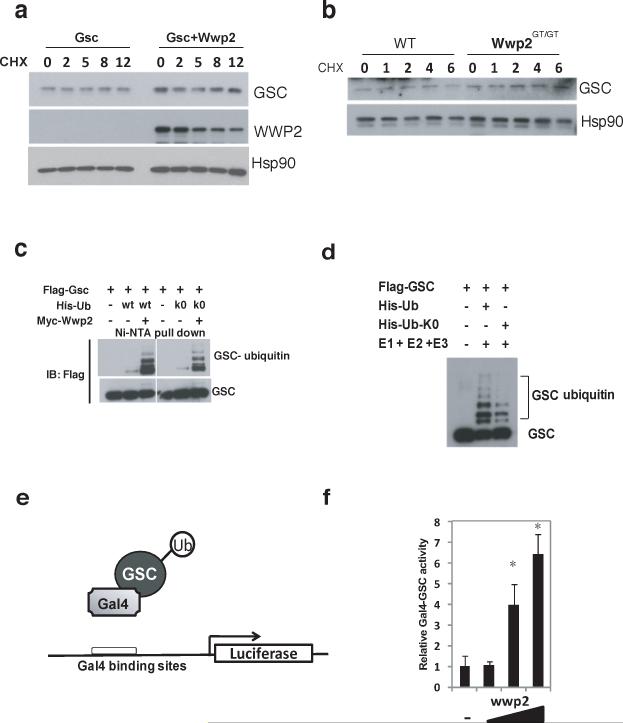
Figure 4. Monoubiquitination augments transcriptional activity of Gsc.
a) Western blot analysis of Gsc and Wwp2 protein levels in cell lysates generated from 293T cells that were transfected with either a Gsc-expression construct or a Gsc-expression constuct and a Wwp2-expression construct. Transfected cells were treated with cyclohexamide (CHX) for 2, 5, 8 and 12 hours prior to cell lysis. b) Endogenous Gsc protein levels were assessed in lysates generated from nasal cartilage cells isolated from WT and Wwp2GT/GT mice by Western blot analysis using anti-Gsc antibody. c) Ubiquitination of Gsc was evaluated in 293T cells transfected with expression constructs of flag-Gsc, myc-Wwp2 and either His-wild type ubiquitin or His-K0-ubiquitin. Ubiquitinated flag-Gsc proteins were detected as described above d) Analysis of Gsc ubiquitination was also assessed using recombinant his-wild type ubiquitin or his-K0-ubiquitin through in vitro ubiquitination assays similar to those described above. e) Schema depicting the Gsc-gal4 fusion protein and reporter construct used in subsequent luciferase experiments. f) Luciferase levels were evaluated following transfection of Gsc-gal4 expresson construct, Gal4-luciferase reporter and increasing amounts of Wwp2 expression construct. Values represent means ± s.d. (n=3) (*p<0.01 compared to empty vector control). Uncropped images of blots are shown in Supplementary Fig.S7.

