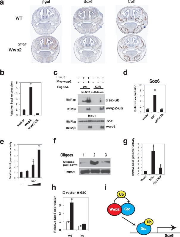
Figure 5. Ubiquitination of Gsc by Wwp2 is required for optimal expression of Sox6.
a) Coronal sections of WT and Wwp2GT/GT skulls were evaluated for expression of β-gal via immunostaining and Sox6 and Col1a1 via in situ hybridization. Scale bar, 1mm b) Sox6 mRNA levels were analyzed by qPCR in WT nasal cartilage cells transduced with a control lentivirus, Wwp2-expressing lentivirus or lentivirus expressing Wwp2 with a non-functional Hect domain (Wwp2-CA). Values represent means ± s.d. (n=3, *p<0.01). c) 293T cells were transfected with expression constructs for flag-myc-Wwp2, His-ubiquitin and flag-tagged Gsc or flag-tagged Gsc-K3R, a Gsc protein with the three lysines mutated to arginine. Ubiquitination of the wild-type Gsc and mutant Gsc-K3R were analysed by immunoprecipitation and Western blot analysis d) wild-type nasal cartilage cells were infected with lentivirus expressing wild-type Gsc or mutant Gsc-K3R or control lentivirus. Transcript levels of Sox6 in these cell populations were then evaluated by qPCR Values represent means ± s.d. (n=3, *p<0.01). e) 293T cells were transfected with Sox6pro-luc reporter construct and increasing amounts of a Gsc-expression construct. Results were normalized to the expression of the pRL-Tk plasmid Values represent means ± s.d. (n=3, (*p<0.01). f) Gsc DNA binding to the Sox6 promoter was determined through nuclear extracts generated from nasal cartilage cells. Gsc binding to region −235 to −185 (probe #1), −187 to −136 (probe #2) and −136 to −87 (probe #3) was detected by Western blotting following oligo pulldown experiments. g) Analysis of Gsc-K3R mutant's ability to transactivate Sox6pro-luc was evaluated posttransfection of 293T cells and was compared to cells transfected with the Sox6pro-luc and construct expressing wild-type Gsc. Levels of luciferase in these experiments were normalized to the expression of the pRL-Tk plasmid. Values represent means ± s.d. (n=3, *p<0.01). h) Sox6 transcript levels were analyzed by qPCR in nasal cartilage cells from WT or Wwp2GT/GT mice infected with control or Gsc-expressing lentivirus. Values represent means ± s.d. (n=3, *p<0.01) i) Model depicting the mechanism through which Wwp2-mediated monoubiquitination of Gsc leads to augmented Sox6 expression. Uncropped images of blots are shown in Supplementary Fig.S7.

