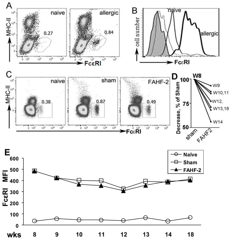
Figure 2. FAHF-2 reduced the number of peripheral blood basophils.
A. Dot plots show an increased percent of basophils in PNA mice, at wk 8 following the last boost, as compared to naïve mice. B. Histogram shows labeled FcεRI expression on cells from allergic (bold line) and naïve mice (thin line). Shaded = unstained cells; gray = isotype controls. C. Percent of basophils in peripheral blood from each group at wk 14 immediately after treatment. D. Reduction of basophil numbers as determined weekly at different time points as indicated. Numbers were normalized to and expressed as % of sham. E. FcεRI MFI of blood basophils from each group over time as in D.

