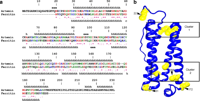
Fig. 1.
Artemin and ferritin structures are related. a The amino acid sequences of artemin (AAL55397) and bullfrog ferritin (P07798) were aligned by CLUSTALW and numbered according to the artemin sequence. *, identical residues; :, conserved substitution; ., semi-conserved substitution. The secondary structure of ferritin was obtained from its crystal structure (1MFR), while the secondary structure of artemin was predicted by the MLRC method at NPS@server. The secondary structures of artemin and ferritin are indicated above and below their sequences. h α-helix, e extended strand, c random coil. b The location of cysteine residues within the artemin monomer was determined by MODELER 7 × 7 using the crystal structure of bullfrog ferritin and residues 21-192 and 2-189 of artemin and ferritin, respectively. Cysteines, yellow. Clustered cysteines are in numbered boxes and modified cysteines are shown by numbered arrowheads. The image was generated with VMD and Raster3D in new cartoon

