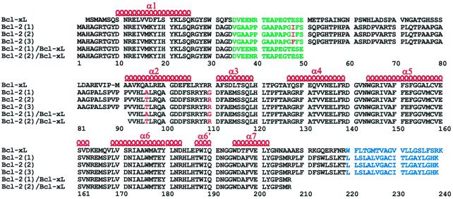
Figure 1.
Sequence alignment of full-length Bcl-xL, the three isoforms of full-length Bcl-2 [denoted Bcl-2(1) (1,2), Bcl-2(2) (3,4), and Bcl-2(3) (5,6)], and the truncated Bcl-2/Bcl-xL chimeras used in this study. Amino acid differences between the Bcl-2 isoforms are shown in red, the truncated loop is shown in green, and the putative membrane-spanning region is shown in blue. α-helices previously identified in Bcl-xL are denoted above the sequence in red.

