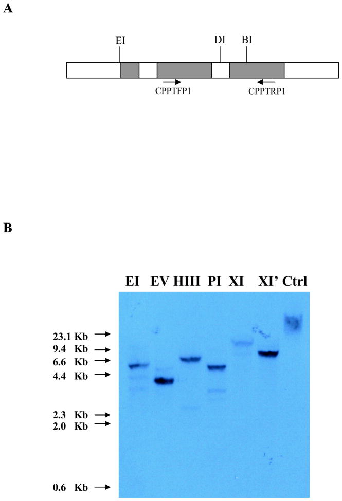
Figure 5.
Schematic diagram showing the structure of Cupip-PTTH gene and Southern blot analysis. A: Cupip-PTTH gene structure and restriction map. Three filled boxes indicate exons, and the open boxes indicate introns. Three restriction enzyme sites are shown as EcoRI (EI), DraI (DI), and BamHI (BI). CPPTFP1 and CPPTRP1 were the primers used for obtaining hybridization probes. B: Southern blot analysis. Ten μg of genomic DNA was digested by 50 units’ restriction endonucleases EcoRI (EI), EcoRV (EV), HindIII (HIII), PstI (PI), XbaI (XI), and XhoI (XI′) respectively; only one major band was observed in each lane. Non-digested DNA was used as a control (Ctrl); sizes of DNA molecular weight markers are shown at left.

