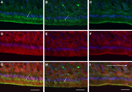
Figure 2.
Immunofluorescent localization of claudin-1 in maturation ameloblasts at higher magnification. Cryosections of rat mandibles were triply labeled with anti-claudin-1 [(A–C), green], rhodamine–phalloidin [(D–F), red] and Hoechst 33342 [(A–I), blue], to co-stain claudin, actin and nuclei respectively. (G), (H), and (I) show merged images of (A,D), (B,E), and (C,F), respectively. The incisal region of an RA band (A,D,G), a part of one SA band (B,E,H), and the apical region of another RA band (C,F,I) are shown. The distal end of the ameloblasts of RAs exhibits both anti-claudin-1 and rhodamine–phalloidin fluorescence (A,C,D,F,G,I). In addition, small round positive fluorescent foci (small arrows) are visible in the supranuclear cytoplasm of the incisal region of the RA and in some of the SA [arrows in (A,B,G,H)]. Bars = 30 μm.

