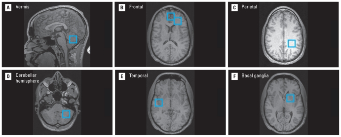
Figure 1.
MR images with blue boxes illustrate the locations of spectroscopy voxels acquired in this study of CLS participants. The seven brain regions included (A) cerebellar vermis, (B) frontal gray matter and frontal white matter (right side of the figure), (C) parietal white matter, (D) cerebellar hemisphere, (E) temporal lobe at the superior temporal gyrus, and (F) basal ganglia. The voxels are acquired in three dimensions with a volume of 8 cc. The figure uses radiological convention for the images, where the left side of the brain appears on the right side of the image.

