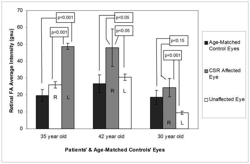
Fig. 1.
Fundus photographs of the affected eyes (A,C,E) and FPF histograms of the affected and unaffected eyes (B,D,F (right, black; left, grey)) of three patients with unilateral CSR. Present were a PED temporal to the left fovea (A), a PED inferotemporal to the right fovea (C), and a blunted foveal reflex with subretinal fluid in the left macula (E). The histograms showed significantly elevated FPF in each CSR-affected eye compared to the contralateral unaffected eye.

