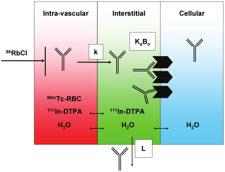
Figure 1. Conceptual illustration of techniques used to measure physiological parameters relevant to antibody uptake in tissues.
The tissue is divided into vascular, interstitial, and cellular compartments (depicted in red, green, and blue, respectively). The blood space (Vv) may be measured using 99mTc-labeled red blood cells (RBC), while the extracellular (i.e. Vv+Vi) space is measured by infusion of 111In-DTPA. The rate of blood flow (Q) to the tissue may be measured as the proportion of a bolus dose of 86RbCl that enters the tissue in a brief time interval. The antibody's receptor, if present, may be expressed on the cell surface, exposed to the interstitial fluid. An antibody in circulation may extravasate from blood into interstitial space at a rate (k), where it may encounter a number (Bo) of receptors for which it has binding affinity (Ka). The antibody may also return to circulation via lymphatic flow (L).

