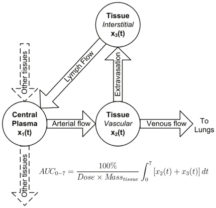
Figure 2. Diagram of physiologically-based pharmacokinetic (PBPK) model to predict antibody uptake in tissues.
Shown is a typical tissue sub-model component of the PBPK model [13] used to assess the influence of parameter variability among literature and measured Vv, Vi and Q values on tissue uptake of an IgG (expressed as AUC0–7). Antibody enters tissue from the central plasma compartment via arterial blood flow where it continues to the lungs via venous blood flow or returns directly to the central plasma compartment through the lymphatic system subsequent to extravasation into interstitial space. The AUC0–7 values listed in Table 4 are the sum of AUCs of absolute antibody amount vs. time in the two tissue compartments (x2 and x3) multiplied by 100% and divided by the product of the total injected dose and mass of tissue, yielding AUC in units of %ID/g × time. Note that the muscle sub-model includes extra compartments, included in the AUC0–7 calculation, that describe FcRn mediated recycling and degradation of antibody.

