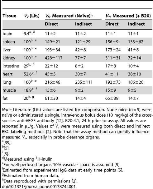
Table 1. Measured vascular volumes (Vv) in naïve and anti-VEGF-administered mice.
Note: Literature (Lit.) values are listed for comparison. Nude mice (n = 5) were naïve or administered a single, intravenous bolus dose (10 mg/kg) of the cross-species anti-VEGF antibody [12], B20-4.1, 24 h prior to assay. All values are reported in µL/g. Values of Vv were measured using both direct and indirect RBC labeling methods [2]. Note that the assay method can greatly influence measured Vv, especially in probe clearance organs.
[39].
[5].
[3].
Measured using 3H-inulin.
For well-perfused organs 10% vascular space is assumed [5].
Estimated from experimental IgG data at early time points [5].
Estimated from human data.
Data reproduced with permissions [2].

