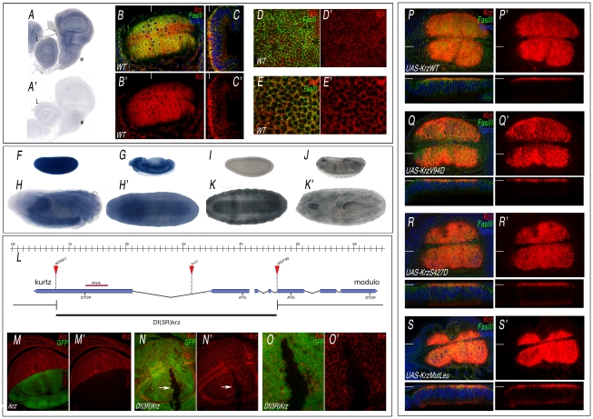
Figure 1. Expression of Krz mRNA and protein.
(A–A′) In situ hybridization with antisense (A) and sense (A′) krz probes in late third instar wing (w) and leg (L) discs. (B–C) Expression of Krz (red), FasIII (green) and Topro (Blue) in the wing pouch of a late third instar disc. (B) Apical focal plane of the wing pouch (B′ is the red channel showing the expression of Krz). (C) Longitudinal sections of the wing pouch at the place indicated in B by a white line. (C′) Red channel of C. (D–E) Transversal sections taken at the apical (D–D′) and medio-lateral (E–E′) level of the wing pouch. D′ and E′ show the expression of Krz (red). (F–H′) In situ hybridization with an antisense krz probe in the blastoderm (F), in stage 13 embryos (G) and in stage 17 embryos oriented laterally (H) or ventrally (H′), showing the prominent expression of krz in the CNS. (I–K′) Expression of Krz protein in the blastoderm (I), in stage 16 embryos (J) and in stage 17 embryos oriented ventrally (K) or dorsally (K′). (L) Representation of the krz gene indicating the intron-exon structure, the position of the ATG and Stop codons, the insertion sites of the e03507, e00739 and krz1 transposons and the extent of the krz deficiency (Df(3R)krz). The scale in Kb is indicated above. (M–M′) Loss of Krz expression (red) in ap-Gal4 UAS-GFP/UAS-ikrz wing discs. The expression of GFP is shown in green. (N–N′) Elimination of Krz expression (red) in clones of cells homozygous for the Df(3R)krz. The clone (arrow) is labelled by the absence of green. (O–O′) Higher magnification of the clone shown in N. (P–S) Expression of Krz (in red), FasIII (in green) and Topro (in blue) in wing imaginal discs expressing the following Krz-FLAG forms in the salEPv-Gal4 domain: UAS-krzWT (P–P′), UAS-krzV94D (Q–Q′), UAS-krzS427D (R–R′), UAS-krzLeu (S–S′). P′–S′ correspond to the red channel of P–S showing the expression of Krz. The planes of each transversal sections (shown below each picture) are indicated by white lines in P–S′.

