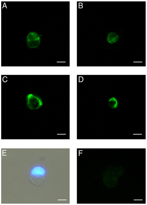
Figure 3. CaSR protein expression and subcellular localization in eUCM-MSCs upon Ca2+- or calcimimetic-induced CaSR stimulation.
Detection of CaSR expression in equine UCM-MSCs in the >8 µm cell line (A, C) and <8 µm cell line (B, D) by immunofluorescence with a primary antibody against a 20 amino acid peptide sequence near the C-terminus of human CaSR and observation by confocal laser scanning microscopy. In both cell lines, cells showing CaSR labeling either predominantly evident whitin the cytoplasm (A, B) or on the plasma membrane (C, D) were present. For each cell, scanning was conducted with 12 optical series from the top to the botton of the cell with a step size of 0.45 µm and images were taken to the equatorial plane. Representative photomicrograph of equine UCM-MSC as observed after thawing, staining with Hoechst 33258 and observed under phase contrast microscopy merged with UV light epifluorescence (E). In this cell, regular round shape morphology and an eccentric nucleus can be seen. Negative minus primary control (F). Scale bar represent 20 µm (A, C, E, F) or 10 µm (B, D).

