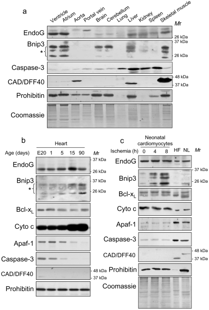
Figure 1. EndoG and Bnip3 are most highly expressed in rat heart, and are located to cardiac myocytes thorough development and during ischemia.
a) Western blot analysis of EndoG, using a new anti-EndoG antibody (AntibodyBcn, BCN4778) and Bnip3 protein expression in total protein extracts from different adult rat tissues. EndoG is detected as a single band (∼27 kDa) and Bnip3 is detected as a group of bands around the 26 kDa mark (asterisk), in agreement with the information provided by the manufacturer. Both proteins are expressed mainly in the heart and skeletal muscle. Detection of EndoG and Bnip3 was performed in two different sets of independent samples with similar results. b) Western blot detection of EndoG, Bnip3 and proteins involved in caspase-dependent cell death in total protein extracts of ventricles from rats of different ages ranging from embryonic day 20 to adulthood. A representative image is shown from three independent experiments. c) Neonatal cardiomyocytes were treated with ischemia and the abundance of EndoG, Bnip3 and proteins involved in the caspase-dependent pathway was analyzed by Western blot in extracts obtained at different time points. Primary heart fibroblasts and neonatal liver samples were added for comparison. A representative image is shown from three independent experiments.

