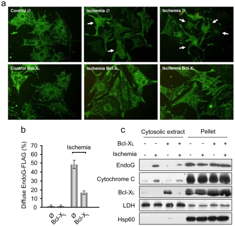
Figure 5. Bcl-xL blocks EndoG translocation in ischemic cardiomyocytes.
a) Immunofluorescence of EndoG-FLAG in control and ischemic (6 hours) cardiomyocytes transduced previously with viruses inducing EndoG-FLAG overexpression and empty viral particles (Ø) or particles inducing Bcl-xL overexpression. Cells were fixed with paraformaldehyde. This procedure allows detection of EndoG-FLAG, even when it is released from mitochondria, but not the mitochondrial marker Hsp60 (see the results section and Supplementary Figure 3). b) Quantification of the experiments described in a). The graph shows the percentage of cardiomyocytes with diffuse pattern of the EndoG-FLAG staining in control and ischemic cultures of empty virus-transduced cells (Ø) and cells transduced with viruses for Bcl-xL overexpression (Bcl-xL), counted in three independent experiments. Error bars are s.e.m. c) EndoG-FLAG release was assessed by Western Blot in cytosolic extracts from control cardiomyocytes (transduced with EndoG-FLAG and empty viruses) and cardiomyocytes overexpressing Bcl-xL (transduced with EndoG-FLAG and Bcl-xL viruses), in standard and ischemic conditions. Expression of Bcl-xL was assessed to confirm its increased expression in cardiomyocytes transduced with Bcl-xL. Lactate dehydrogenase (LDH) expression was checked as a cytosolic marker and Hsp60 expression was assessed as a mitochondrial membrane marker. The image shows representative results from 3 independent experiments.

