Figure 1.
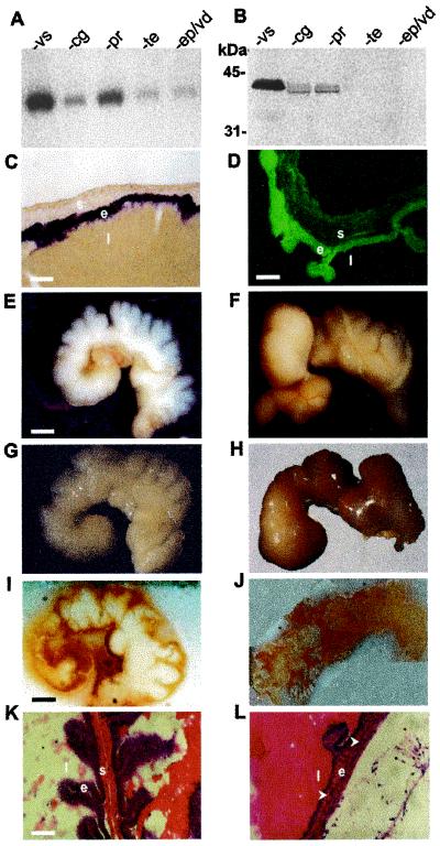
PN-1 expression in the wild-type male reproductive tract and seminal gland morphological phenotypes in PN-1−/− males. (A) PN-1 transcript and (B) PN-1 protein contents of the seminal vesicle (vs), the coagulating gland (cg), the prostate (pr), the testis (te), and the epididymis-vas deferens (ep/vd) of adult wild-type mice. (C) PN-1 in situ hybridization and (D) PN-1 immunocytochemistry in the adult seminal vesicle. PN-1 mRNA is expressed in the secretory epithelial cells (C); PN-1 protein is predominantly found at the apical side of these cells (D) as well as secreted into the lumen. e, epithelium; l, lumen; s, stroma. (E–H) Morphology of seminal vesicles from 4- and 10-month-old PN-1+/+ mice and PN-1−/− mice. At 4 months, the seminal vesicles of the mutant show a reduction in the number and the depth of the folds of the stromal sheath, obstruction, and/or dilatation of the distal part of the gland and a yellowish tint in the secretory fluid (F compared with E). At 10 months, the phenotype was more severe: these organs showed a massive dilatation and the secretory fluid showed a strong yellow-brownish coloration (H compared with G). (I and J) Compared with the homogenous white appearance of the wild-type seminal vesicle (I) a micro granular-like-structure with a brownish color was detected in the mutant at 6 months (J). (K and L) Histology of seminal vesicle epithelium from 2-month-old (K) and 10-month-old (L) PN-1−/− mice. Frozen sections were stained with hematoxylin and eosin. Note the decreased organization of the epithelium layer and the apparent loss of the stroma in 10-month-old mutant mice (arrows). [Scale bars: (C and K) 50 μm; (D) 35 μm; (E) 1.5 mm; (I) 1.2 mm.]

