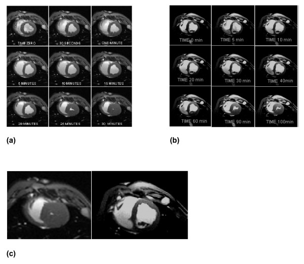
Figure 1.
Cardiac Magnetic Resonance images of the swine heart at the level of the mid-ventricular short axis view of the right and left ventricles. a) A representative normothermia animal over 30 minutes of untreated ventricular fibrillation. Stone heart in this example developed at ~25 minutes; b) A representative hypothermia animal over 100 minutes of untreated VF. Stone heart in this animal developed at ~90 minutes. Time 0 refers to time ventricular fibrillation (VF) was induced, and next 8 views were at respectively labeled duration of untreated VF; c) MRI "zoom" comparison of the 30 minute image for the representative normothermia animal (left) and the hypothermia animal (right).

