
Figure 1.
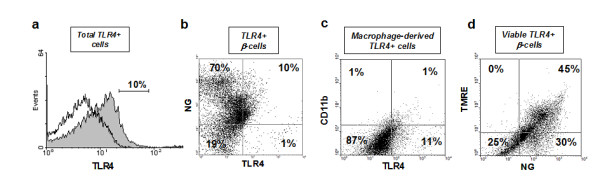
Flow cytometric analysis of TLR4 expression in human islet cells. Free-floating 8-12 day islet cultures (500-1000 IEQ) were dissociated into single-cell suspensions and assessed by flow cytometry. A representative image from three independent experiments is shown. (a) Histogram compares total TLR4-positive cells (gray) with isotype-matched goat anti-rabbit IgG control for TLR4 staining (no color). (b) The right upper quadrant of the dot plot shows the TLR4 positive β-cell population, double-stained for Newport Green (NG) and TLR4-APC conjugate, (c) The right upper quadrant of the dot plot shows TLR4-positive macrophage-derived cells (non β-cells) double stained with TLR4-APC conjugate and CD11b-PE conjugate, (d) Tetramethylrhodamine (TMRE) and NG double staining marks viable β-cells.