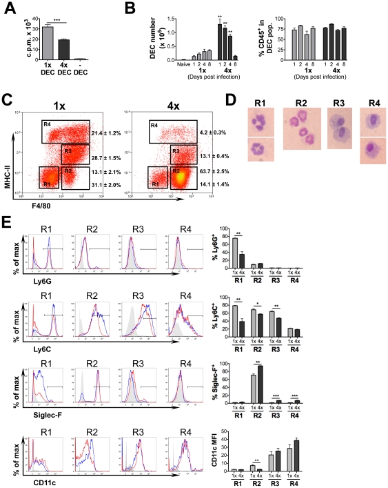
Figure 3. Dermal exudate cells (DEC) from 4x mice are inefficient at supporting antigen-specific CD4+ cell proliferation and comprise a large influx of eosinophils but a reduction in MHC-II+ cells.
(A) DEC recovered from cultured biopsies of 1x and 4x infected skin were co-cultured with purified CD4+ T cells from the sdLN of 1x mice and stimulated with parasite antigen. Bars show the mean c.p.m. + SEM (n = 5 DEC samples) and is representative of 4 experiments performed with similar results. (B) Numbers of DEC recovered from naïve, 1x and 4x mice, and the proportion which are CD45+ (mean + SEM, n = 6 pinnae/time point). (C) Representative flow cytometry dot plots of DEC recovered on day 4 labelled for MHC-II and F4/80. Values show mean percentage ± SEM of the gated populations R1-R4 (n = 6 mice). (D) Morphology of DEC sorted by MoFlo into R1-R4 on the basis of F4/80 and MHC-II stained with DiffQuick. (E) Representative flow cytometry histogram plots of R1-R4 cell populations labelled with antibodies against Ly6G, Ly6C, SiglecF, and CD11c from 1x (blue) and 4x (red) mice; solid grey plot shows the extent of isotype control antibody staining. Also shown is a bar chart for each marker showing the mean values + SEM for 5 individual mice. Data is representative of at least two experiments.

