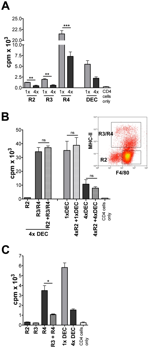
Figure 5. DEC from 4x mice include suppressive and functionally impaired MHC-II+ cells, but eosinophils do not directly cause cell hypo-responsiveness.
(A) R2, R3 and R4 cells (1×10⁴) from 1x and 4x DEC were co-cultured with purified CD4+ cells from 1x sdLN in the presence of parasite antigen. (B) R2 eosinophils (2×10⁴) from 4x DEC were co-cultured with purified CD4+ cells from 1x sdLN in the presence of parasite antigen, or together with mixed R3/R4 cells, or unsorted 1x and 4x DEC populations (all 2×10⁴). Bars show CD4+ cell proliferation as mean c.p.m. + SEM (n = 5). (C) Sorted R3 and R4 cells from 4x DEC were cultured separately, or combined, with purified CD4+ T cells from sdLN of 1x mice. Significances are shown between groups indicated by connector bars. Sorted DEC fractions were pooled from 15–35 mice and bars are mean + SEM of five replicate wells and are representative of 2–3 experiments.

