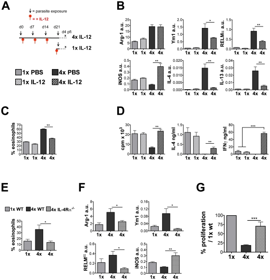
Figure 7. Treatment of 4x mice with rIL-12, or multiple infection of IL-4Rα−/− mice reduces eosinophilia and restores the lymphocyte responsiveness in the sdLN.
(A) Treatment regime of rIL-12 administration to 4x mice 2 days after the 1st, 2nd and 3rd infection, and 2 days prior to infection for 1x mice. (B) Transcript analysis by qRT-PCR of DEC from PBS or rIL-12 treated 1x and 4x infected mice expressed in arbitrary units (a.u.) relative to GAPDH shown as mean + SEM (n = 4–5). (C) Mean percentage of cells in R2 DEC recovered from rIL-12 or PBS treated 1x and 4x mice + SEM (n = 4–5 mice). (D) Antigen-specific in vitro proliferation and cytokine production by sdLN cells from PBS or rIL-12 treated 1x and 4x mice. Results show the mean c.p.m., or pg/ng cytokine/ml, + SEM. (E) Percentage of Siglec-F+ and F4/80+ cells in DEC recovered from 1x WT, 4x WT and 4x IL-4Rα−/− mice. Bars show mean percentage + SEM of the relevant gated region (n = 5 mice). (F) Transcript analysis of Arg-1, RELMα, Ym-1, and iNOS genes in the total DEC performed by qRT-PCR (n = 5). (G) Antigen-specific in vitro proliferation by sdLN cells. Results are shown as the mean percentage change compared to the level of proliferation generated by 1x WT cells + SEM (n = 9–16).

