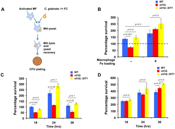
Figure 3. Macrophage Fe status determines the degree of Sit1 dependence for C. glabrata to survive macrophage killing.
A Schematic flowchart of the macrophage infection assay developed for C. glabrata virulence studies. C. glabrata cells were grown in low Fe medium for 5 hours and pulsed with 10 µM ferrichrome (FC) for 30–45 minutes prior to exposure to activated mouse macrophages. C. glabrata cells were recovered from the lysed macrophages, plated on YPD and colony forming units (CFU) counted after 2 days at 30 °C. Data were analyzed with use of one-way ANOVA. B Sit1-mediated siderophore utilization enhances the survival of C. glabrata to macrophage killing activities (left columns) and this is ameliorated by elevated intracellular macrophage Fe. Macrophages were Fe-loaded with ferric ammonium citrate [20 µM] for 2 days prior to infection with C. glabrata. Each experiment included 6 replicates per experimental sample. Results are shown as mean survival compared to the wild type grown in the absence of ferrichrome supplementation (dashed line) and are representative of at least 3 experiments. Bars represent standard error. C The human monocytic cell line, U937, was differentiated into mature macrophages prior to infection with the indicated C. glabrata strains grown under Fe deficiency without or with supplementation of 10 µM FC and yeast survival evaluated after 18, 24 and 36 hours of infection. The results depicted show the mean survival of the wild type, sit1Δ and sit1Δ::SIT1 strains grown in the presence of FC prior to macrophage infection. Bars indicate standard error. D U937 macrophages were Fe-loaded with ferric ammonium citrate [20 µM] for 2 days prior to infection with C. glabrata strains grown under Fe deficiency without or with supplementation of 10 µM FC and yeast survival evaluated after 18, 24 and 36 hours of infection, as described above.

