Fig. 4.
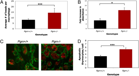
Loss of progranulin accelerates phagocytosis in mouse macrophages. Peritoneal macrophages were cultured from Pgrn+/+ and Pgrn−/− animals and incubated with FITC-labeled polystyrene beads (A), yeast cell-wall particles (zymosan) (B), or pHrodo-labeled apoptotic thymocytes (C and D) in the presence of 10% heat-inactivated FBS. (A) The average number of beads engulfed by each macrophage after 90 min incubation was determined (n ≥ 100 cells per condition). Error bars represent SD (Student t test, ***P < 0.0001). (B) Average increase in zymosan uptake by colorimetric assay after 90 min of incubation. Error bars represent SEM of two independent experiments (Student t test, *P < 0.05). (C and D) The average number of apoptotic thymocytes engulfed by each macrophage after 90 min of incubation was determined. Representative fields are shown in C (with pHrodo-labeled apoptotic thymocytes in red and CD-11b-FITC–labeled macrophage membranes in green) and average counts shown in D (n ≥ 100 cells per condition). Error bars represent SEM (Student t test, ***P < 0.0001).

