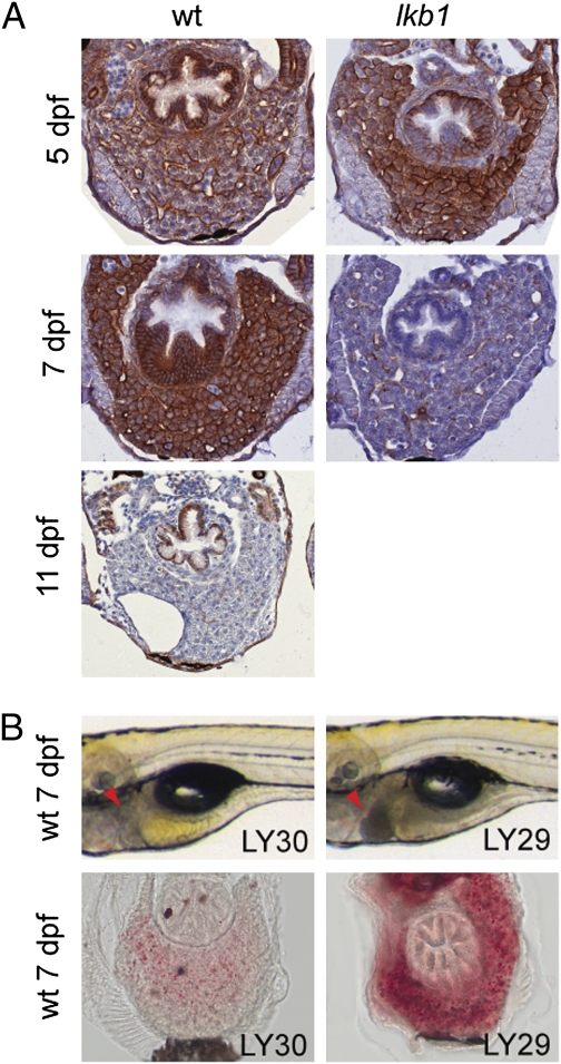
Fig. 5.
Deregulation of PI3K signaling in lkb1 mutants. (A) Transverse sections of WT and lkb1-mutant livers at indicated days of development stained with an antibody against phospho-AKT. Strong phospho-AKT staining is detected in WT and lkb1-mutant livers at 5 dpf. WT liver is strongly stained at 7 dpf, whereas phospho-AKT staining is barely detectable in 7-dpf lkb1-mutant liver and in starved WT at 11 dpf. (B) Inhibition of PI3K signaling leads to a starvation-like phenotype in WT larvae. WT larvae at 7 dpf treated for 3 d with either LY29 or its inactive analog LY30. LY29 treatment of WT larvae at 4 dpf leads to dark liver (arrowheads) and abnormal hepatic steatosis as revealed by ORO staining. Treatment with the inactive analog LY30 has no effect in the morphology of the larvae.

