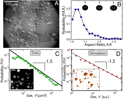
Fig. 1.
Size and shape of X. laevis oocyte nucleoli. (A) DIC image of nucleoli in the X. laevis GV. Nuclear bodies can be readily seen in DIC. Most of these are extrachromosomal nucleoli. (B) We plot the distribution of nucleolar aspect ratios obtained from analysis of GFP∷NPM1 images of nucleoli (Inset in C). The average is 1.07 ± 0.06; a perfect sphere would be 1.0. Thus, most nucleoli are highly spherical. (C) Distribution of nucleolar volume exhibits a power-law distribution with an exponent of -1.5. Inset shows an image of GFP∷NPM1–labeled nucleoli (scale bar, 20 μm). (D) Monte Carlo simulation of fusing droplets, with a slow constant influx of small droplets. The distribution of droplet volume exhibits power-law behavior with an exponent of -1.5. A snapshot of a subregion of the simulation is shown in the Inset.

