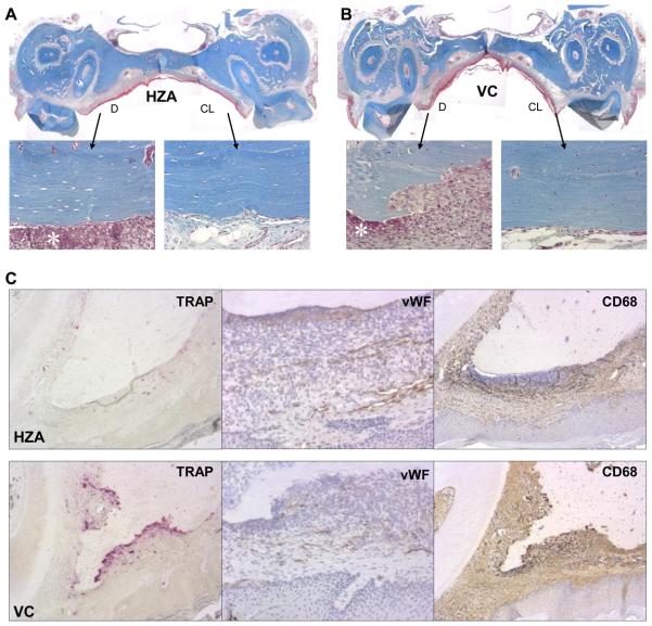
Fig. 4.
Wound healing in the palate. The palatal alveolar bone was denuded 12 weeks after the initiation of zoledronate treatment (see Figure 1). Healing at 2 weeks post-op was evaluated. Representative photomicrographs of Masson's trichrome-stained sections of the palates of rats treated with high-dose zoledronate (A) and vehicle control (B) (40x). Epithelialization of wounds was observed in most cases. The denuded (D) and contralateral intact (CL) sides are magnified (200x) to show osteocyte lacunae. Neutrophil aggregation (*) was noted next to the unresorbed bone surfaces. C: Histological staining for osteoclasts (TRAP) (100x), von Willebrand factor positive endothelial cells (vWF) (200x), and macrophages/monocytes (CD68) (100x). TRAP+ cells are shown in red color and vWF+ cells and CD68+ cells in brown color. The denuded alveolar bone areas in the HZA (top row) and in the VC (bottom row) are shown.

