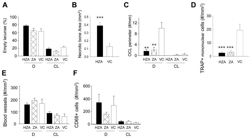
Fig. 5.
Histomorphometric assessement of wound healing. A: The percentage of empty osteocyte lacunae in the predefined AOI in the palate was determined histomorphometrically. B: The entire necrotic bone area was histomorphometrically measured and compared between HZA and VC. C: Osteoclast numbers per linear perimeter were measured in the AOI. In the denuded site significant decrease was noted in HZA and ZA compared to control rats. D: The number of TRAP+ mononuclear cells per mm2 was determined in the connective tissue area in the denuded site. Significant reduction in preosteoclast numbers was found in HZA and ZA compared to control rats. The numbers of vWF positive blood vessels (E) and CD68 positive cells (F) were assessed in the connective tissue. Data are shown as mean ± SE (n=8/group). **p < 0.01, ***p < 0.001 by one-way ANOVA.

