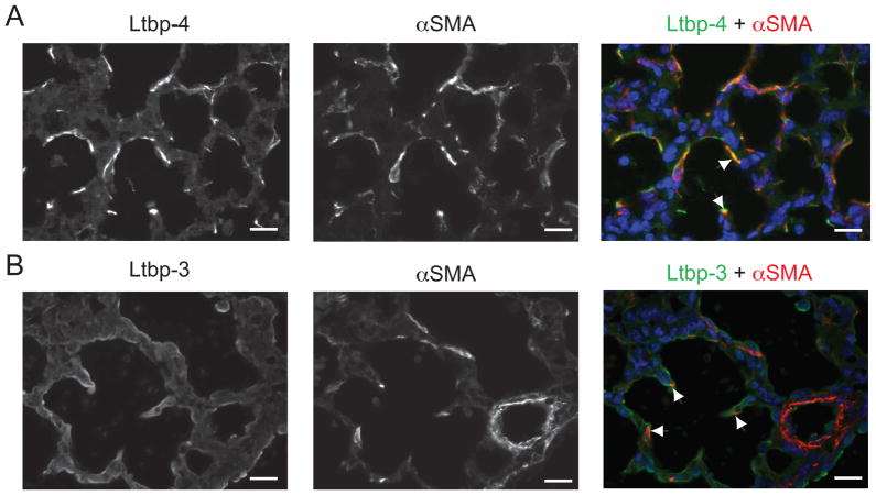
Figure 3.
Lung myofibroblasts synthesize both Ltbp-3 and Ltbp-4. P0.5 WT lung sections were analyzed by immunofluorescence using antibodies against SMC/myofibroblast specific αSMA and an antibody against either Ltbp-4 (A) or Ltbp-3 (B). Nuclei were stained with DAPI. A. Double immunofluorescence of WT cells stained with both Ltbp-4 (left panel) and αSMA (middle panel). The yellow signal indicates overlap of Ltbp-4 and αSMA signals (right panel, arrowheads). B. Double immunofluorescence of WT cells stained with both Ltbp-3 (left panel) and αSMA (middle panel). A small but real number ofαSMA-positive cells (arrows) was also stained with Ltbp-3 (arrowheads), indicating that Ltbp-3 is expressed in some lung myofibroblasts. Bars: 20μm.

