Figure 4.
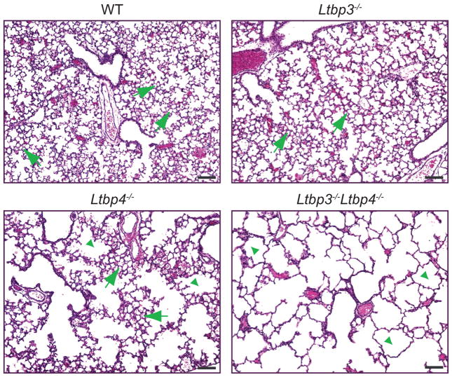
Histology of Ltbp4S−/− and Ltbp3−/−;Ltbp4S−/− lungs at P7. Lung sections were stained with hematoxylin and eosin. WT and Ltbp3−/− lungs display normal morphology with small terminal air sacs (arrows) generated by lung septation and alveolarization after birth. In Ltbp4S−/− lungs some areas undergo alveolarization, and have clusters of small terminal air sacs (arrows). Those areas are separated by areas with large air-spaces, where lung septation is arrested (arrowheads), giving Ltbp4S−/− lungs a patch like appearance in tissue sections. In Ltbp3−/−;Ltbp4S−/−lungs all terminal air sacs are greatly enlarged (arrowheads) indicating a severe decrease in lung septation after birth. Bars: 100 μm.

