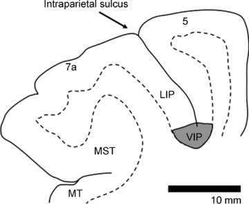
Figure 1. Location of the ventral intraparietal area (area VIP) in the depth of the intraparietal sulcus.

Borders between grey and white matter are based on histological sections. Neighbouring areas 7a, LIP (lateral intraparietal area) and 5 are indicated.
