
Figure 5. Retinal flow fields and related neuronal responses.
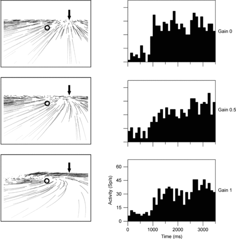
The left columns show the retinal flow fields seen by an observer moving in a rightward heading direction on top of a ground plane. All three panels display the same heading (arrow) but differ in terms of simulated eye movements. The pattern as shown in the top panel occurs in the absence of eye movements (Gain = 0.0). In the middle and bottom panel, eye movements which spontaneously occur in primates are included (Gain = 0.5 (middle) and Gain = 1.0 (bottom)). These eye movements track the motion in the direction of gaze (circle) and distort the structure of the flow on the retina, generating a motion pattern that resembles a spiral and in which the motion in gaze direction (circle) is minimized. Invariant responses to heading should be the same in all three cases, since heading is identical although flow structure is different. The right column shows responses from a neuron responding to rightward heading irrespective of eye movements. Histograms show the neuron's firing rate over time during presentation of optic flow stimuli.