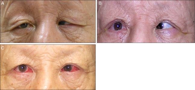
Fig. 3.
(A) Preoperative photography shows both upper and right lower eyelid entropion. An overactive frontalis muscle compensates for both blepharoptosis and dermatochalasis. (B) Photography after surgical eyelid correction. Frontalis muscle overactivity is relieved. (C) Photograph obtained on the 7th day after correction of esotropia. Slight esodeviation remains, and subconjunctival hemorrhage is present.

